Skin tumors
Several types of skin lesions need to be surgically removed from the face:
- Benign lesions when they are unsightly
- Precancerous lesions
- Cancerous lesions
Skin cancers are the most common type of cancer, and tend to occur on the face, as this area is particularly exposed to the sun. There are over 100,000 new cases of skin cancer in France every year.
Schematically, there are 3 types of facial skin cancer and nasal cancer:
- Basal cell carcinoma is the most common and the least serious.
- Squamous cell carcinoma (formerly known as squamous cell carcinoma)
- Malignant melanomas are rarer skin cancers (4,000 to 5,000 new cases per year in France), but more serious, requiring medical and surgical treatment in specific centers.
Predisposing factors
- Sun: excessive sun exposure, especially in patients with white skin
- Exposure to ionizing radiation
- Exposure to certain chemicals (arsenic, tar)
- Trauma and chronic wounds
- Exposure to certain viruses such as papilloma
- Trauma or chronic wounds
- Certain drugs, particularly immunosuppressants, in transplant patients
Secondly, the frequency of these tumors increases with age, and runs in families, certainly via the skin phototype.
Patients who have already suffered from this type of lesion should be closely monitored by their dermatologist, as they are at much greater risk of developing others.
Finally, there are precancerous skin lesions that need to be detected and treated before they degenerate into full-blown skin cancer.
- actinic keratoses
- Bowen's disease
- papillomatoses, xeroderma pigmentosum, basal cell nevomatosis...).
- radiodermatitis (skin lesions caused by radiotherapy)
- burn scars and other scars
- chronic wounds
- lichen and leukoplakia

.jpg)
.jpg)
Pre-epitheliomatous keratosis Seborrheic keratosis with areas of degeneration
.jpg)


BOWEN's disease of the temple Degenerated preauricular keratosis Non-degenerated pigmented acanthosis seborrheic wart
Diagnosis: nasal and facial cancers
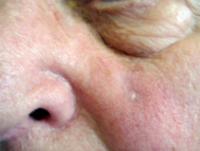
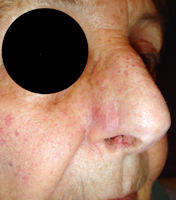
BASAL CELL CARCINOMAS
Three times out of four, they are located on the head and neck. Common locations are the face (nose, eyelids, lips, cheeks, forehead, temples), scalp and upper trunk, but they can occur in other areas of the body. It does not usually occur on a precancerous lesion.
They can take the form of a raised red or pinkish pimple, a small red or white spot with a raised border, or a sore that won't heal.
Basal cell carcinoma does not metastasize, but has the potential to be locally invasive, leading to significant tissue destruction. There are three clinical varieties:
- nodular basal cell carcinoma: a firm, well-limited, smooth tumor that can simulate a cystic lesion or spread centrifugally. This is the most common form.
- superficial basal cell carcinoma: an erythematous, scaly plaque, bordered by pearls that are sometimes barely visible to the naked eye and spreading progressively. It occurs mainly on the trunk, and is sometimes initially multifocal.
- Scleroderma basal cell carcinoma: takes the form of a whitish, poorly defined, sometimes atrophic scar. This is the most dangerous form
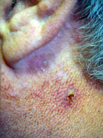
SQUAMOUS CELL CARCINOMAS
They often occur on sun-exposed areas (head and neck, décolleté, upper body, forearms, hands and nails), but can also affect other areas such as the mouth or genitals. On sun-exposed skin, they often begin as a white crust (keratosis) that gradually thickens and ulcerates to form an irregular, raised-edge sore.
Typical examples include :
- a crusty, yellowish, indurated lesion with central ulceration (figs. 23.5 and 23.6).
- a vegetative or budding lesion
- or a combination of the two
In all cases, beware of pimples, scabs, spots that persist and change, or wounds that don't heal quickly or bleed regularly.
In case of doubt, a skin biopsy will confirm the diagnosis.
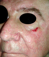
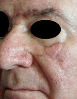
Examples of basal cell carcinomas
.jpg)
.jpg)
.jpg)
Cheek BCC Nose BCC Scleroderma BCC Advanced BCC
Examples of cutaneous squamous cell carcinomas (squamous cell carcinomas)
.jpg)
.jpg)
CE of the neck ulcer CE of the cranial vault CE on a frontal bump
Treatment principles
Treatment is first and foremost preventive: primary prevention by avoiding exposure to the sun, and secondary prevention through regular check-ups if you've already had such a lesion (approximately every year).
Treatment of the lesion is above all its local destruction, notably by surgical exeresis.
Avoiding sun exposure is the best way to prevent skin cancer. Exposure to the sun and ultraviolet radiation are the main risk factors for skin carcinoma. Sunbathing should therefore be avoided, as should exposure to the sun during the hours when solar radiation is most intense (from 11 a.m. to 4 p.m.). A protective sunscreen (at least SPF 20) should be applied every two hours, and protective clothing and a hat should be worn after every bath.
Treatment of nasal and facial cancers most often involves surgery. The lesion must be removed with a safety margin that varies according to the type of carcinoma and its stage of development.
Depending on the type of lesion and its location, surgery ranges from simple outpatient excision and suturing to two-stage excision with plastic reconstruction under general anesthesia.
Microscopic analysis of the tumour after removal ensures that it has been completely removed. Some squamous cell carcinomas may require removal of neighbouring lymph nodes.
Electrocoagulation, the C02 laser, the application of liquid nitrogen, radiotherapy, the application of an immunomodulating cream (imiquimod or aldara®) or, more recently, photodynamic therapy are sometimes used to treat certain superficial basal cell carcinomas, but these methods have the disadvantage of not allowing analysis of the treated lesion. Pre-cancerous keratoses (actinic keratoses) can be treated with nitrogen, fluorouracil cream, electrocoagulation, the C02 laser, photodynamic therapy, or sometimes surgery.
Examples of surgical results
Basal cell carcinoma of the nose wing
.jpg)
.jpg)
Preoperative appearance resection-closure with flap Postoperative appearance
Basal cell carcinoma of the nostril sill
.jpg)
.jpg)
.jpg)
Preoperative appearance resection-closure with island flap Postoperative appearance
Double basal cell carcinoma of the cheek and nose wing
.jpg)
.jpg)
.jpg)
Pre-op resection-closure with double flap Post-op appearance
advancement and island
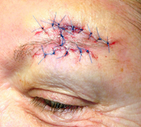
Basal cell carcinoma of the eyebrow
.jpg)
.jpg)
.jpg)
Preoperative appearance Resection and flap alignment Resection-closure with flap Postoperative appearance
Basal cell carcinoma of the lower eyelid
Preoperative appearance Postoperative appearance
Jugal squamous cell carcinoma


Preoperative appearance Resection and flap tracing Postoperative appearance
